


What is a mammogram?
A mammogram is a specialised X-ray (radiograph) of the breast. It is a safe procedure that uses a very low dose of radiation to show breast tissue in detail.
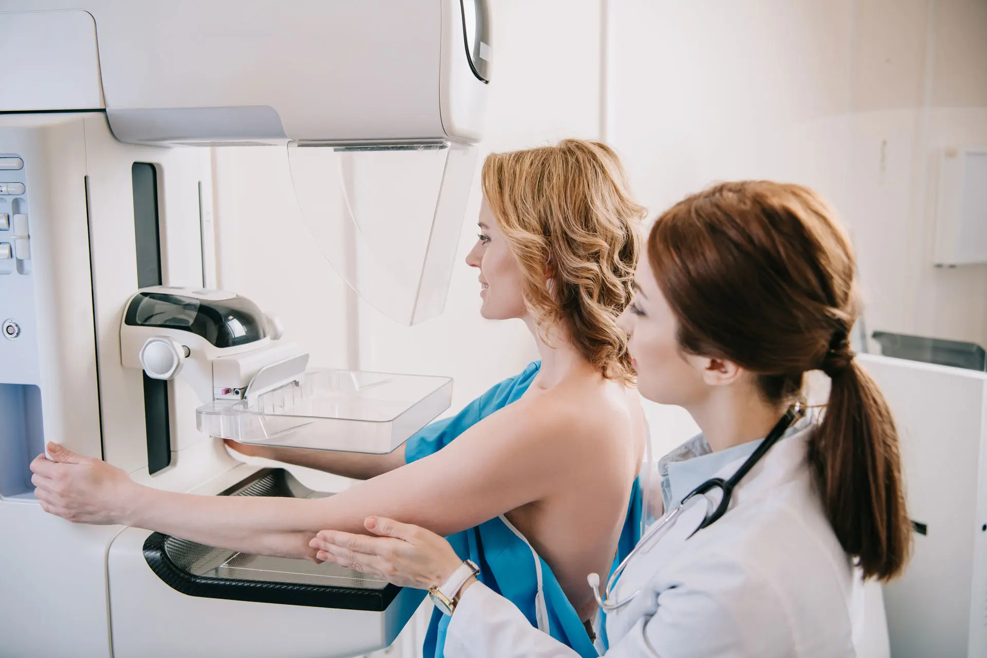
Each breast is imaged separately. During the scan, the breast is held gently but firmly between two plates for a few seconds while images are taken.
Two X-ray views are taken:
- From above (top to bottom)
- From the side

What is mammography used for?
Mammography is used in two key ways:
Breast screening mammography
- A routine wellness check using breast X-rays
- Can detect breast changes before symptoms are present
- May identify abnormalities up to two years before a lump can be felt
Breast diagnostic mammography
Used when there is a breast concern such as:
- A lump
- Thickening
- Pain
Early detection of small breast cancers is important, as it allows treatment to begin sooner.
We are proud to be the 1st Breast Care Clinic in Auckland to offer 3D Breast Tomosynthesis.

What is 3D mammography (breast tomosynthesis)?
A 3D mammogram, also known as breast tomosynthesis, is an advanced form of mammography.
It uses X-ray technology to take multiple images of thin slices of the breast, which are then reconstructed into a three-dimensional image.

Why is 3D tomosynthesis important?
In standard mammography:
- Glandular and fibrous tissue appears white
- Fat appears black
- Cancer can also appear white and may be harder to detect, especially in dense breasts
With 3D tomosynthesis:
- Images are taken through the breast during the mammogram
- The computer reconstructs these into 1mm slices
- These are reviewed alongside 2D digital images
Benefits include:
- Improved cancer detection
- Reduced false negative results
- Greater accuracy in dense breast tissue
- Fewer unnecessary biopsies
Who should have a mammogram?
Regular screening mammography is recommended for all women over 40 years of age.
Additional recommendations include:
- Women aged 40–50 years should have an annual mammogram
- Women with a previous history of breast cancer should have annual mammography
- Women with a first-degree relative (for example, a mother or sister) who has had breast cancer may begin mammograms 10 years earlier than the age at which their relative was diagnosed
Your healthcare provider will guide you on the most appropriate screening schedule based on your individual risk.
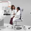
What to expect during your mammogram
- Each breast is positioned on the imaging machine (one at a time)
- Gentle compression is applied briefly
- Images are taken within a few seconds
- The process is quick and safe

Mammograms during pregnancy and breastfeeding
Mammography and ultrasound can be safely performed during pregnancy and breastfeeding when clinically required. Your healthcare team will recommend the most appropriate imaging approach based on your individual situation.
Hormonal changes may increase breast density during this time, which can affect imaging clarity. For this reason, ultrasound is often used alongside mammography to provide a more comprehensive assessment.
If you notice any new breast changes, such as a lump or persistent discomfort, it is important to seek medical advice.
Learn more about breast imaging during pregnancy and breastfeeding
You will be asked to remove your clothing from the waist up, and given a gown to wear. For ease of undressing, you may like to wear a shirt and a skirt, or trousers.
Please do not wear deodorant or talcum powder on your breasts or armpits on the day of your examination. We do however, provide wipes and deodorant for your personal use.
Discuss any breast symptoms or problems with the mammographer before the examination.
It is important for the radiologists interpreting the mammogram to have previous studies for comparison as they compare previous images with new images to look for any areas of change to the breast.
Please remember to also bring the referral note from your doctor.
A skilled mammographer will take at least two pictures of each breast. Each breast is placed on the machine and compressed with a plastic paddle. This flattening of the breast will reduce the breast thickness and hold the breast still, which optimises image quality and reduces radiation dosage.
Most women would describe the compression as uncomfortable, rather than painful. If you have found the compression painful, please discuss your concerns with the mammographer.
Sometimes further images may be needed to show an area of the breast more clearly. Don't be concerned if this occurs. An ultrasound scan may also be necessary to complete your examination.
In most instances having a mammogram only takes about 20 minutes. We may in some cases also need to perform an ultrasound.
After the images are taken of the breast, you will be asked to wait until the mammographer has viewed them to see if any more are needed.
The Radiologist may ask if you wish to see the images and will give you your result then.